Prof. LIANG Gaolin’s team from University of Science and Technology of China (USTC) recently developed a method of persistent bioluminescence (BL) imaging of tumor and verifies it in vivo of tumor-bearing mouse model, by making use of intracellular self-assembly of cyclic D-luciferin-based nanoparticles.
This new method enables long-term tracing of fatty acid amide hydrolase (FAAH) activity. FAAH is a kind of serine hydrolase enzyme and it can degrade anandamide to keep nerve systems in order. Detecting its activity in a long period is very important, because its overexpression results in serious chronic disorders, such as inflammation, anxiety, and cannabinoid dependence.
Long-term detection of FAAH activity has been challenging. One time-and-energy-consuming and not direct method of evaluating FAAH activity is to take the tissue of the mice out, homogenate and then analyze the hydrolyzed products by FAAH. Another method is positron emission tomography (PET), which can get and analyze the image of FAAH activity directly, but the half-life period of PET tracer is no more than 2 hours. BL imaging has also been adopted in detection of FAAH activity, but the half-life period of luciferin is less than 30 minutes, and no more than 5 hours even with nanoparticles.
To improve the current BL imaging method, the research team led by Professor LIANG Gaolin employed the self-developed “smart” strategy of self-assembly/disassembly (Angew. Chem. Int. Ed. 2015, 54, 9700-9704;ACS Nano 2015, 9, 761-768;ACS Nano 2015, 9, 5117-5124), rationally designed a latent luciferin micromolecule 1. 1 assembled into luciferin nanoparticle immediately after entering the cell, then the nanoparticle disassembled and released luciferin gradually, resulting in long-term period imaging of FAAH activity, as depicted in the following figure. Animal experiment showed the method can obtain the continuous image of FAAH in tumor for as long as 2.5 days, without being toxic to mouse model. The probe 1 is expected to be used in clinic to screen FAAH inhibitor in vivo and diagnose related diseases.
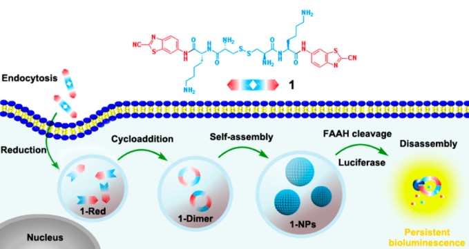
Figure. Chemical structure of 1 and schematic illustration of intracellular reduction-controlled self-assembly and FAAH-directed disassembly of cyclic D-luciferin-based 1-NPs for persistent bioluminescence imaging of FAAH. (Credit: Prof. LIANG Gaolin)
The research observation has been published online in ACS-Nano online,entitled “Intracellular Self-Assembly of Cyclic D-Luciferin Nanoparticles for Persistent Bioluminescence Imaging of Fatty Acid Amide Hydrolase” (DOI: 10.1021/acsnano.6b03412). The article is first-co-authored by post doctor YUAN Yue from the School of Chemistry and Materials Science at USTC and Doctor WANG Fuqiang from Nanjing Medical University. The corresponding author is Professor LIANG Gaolin from USTC.
The research has been sponsored by Collaborative Innovation Center of Suzhou Nano Scienceand Technology, the Major Program of Development Foundation of Hefei Center for Physical Science and Technology, Hefei Science Center CAS, the China Postdoctoral Science Foundation, the Fundamental Research Funds for the Central Universities, and the National Natural Science Foundation of China.
The link of the article: http://pubs.acs.org/doi/abs/10.1021/acsnano.6b03412
Corresponding author information:
Prof. LIANG Gaolin,
gliang@ustcnet., http://en.scms.ustcnet./faculty/professors/201107/t20110703_115182.html.